3175x175(CURRENT).thumb.jpg.b05acc060982b36f5891ba728e6d953c.jpg)
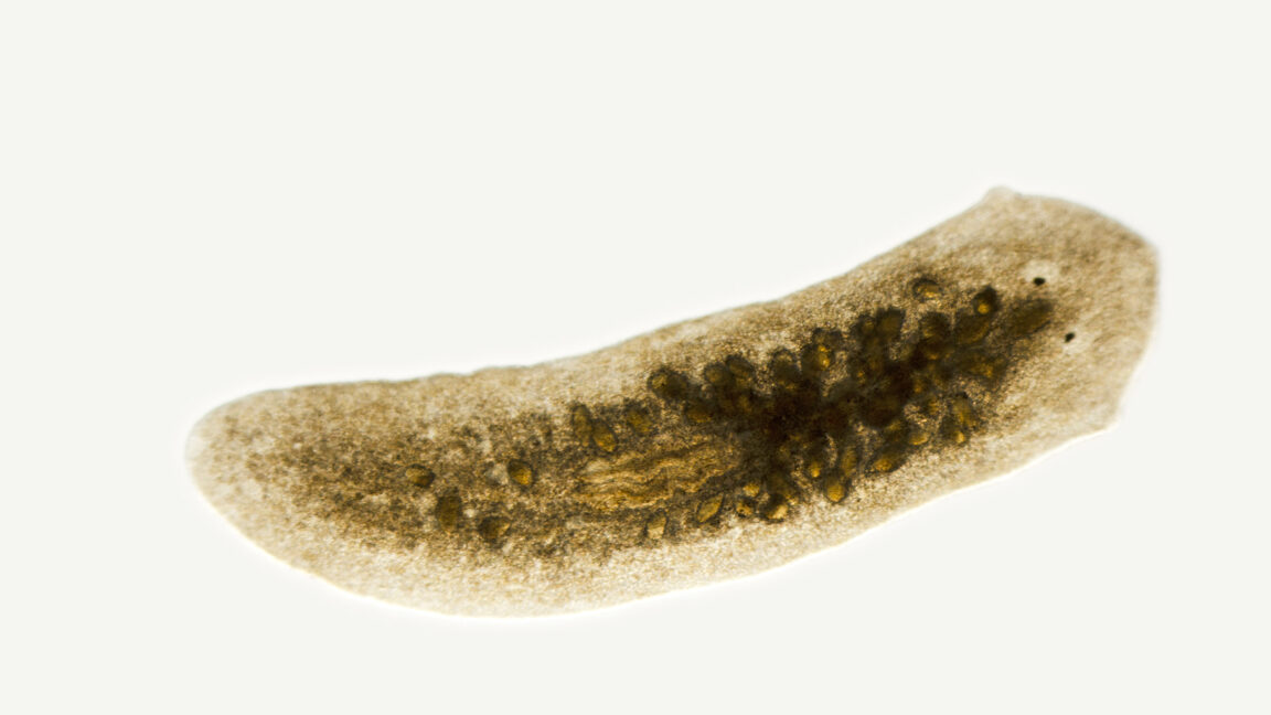
Planaria can't replace a missing head until after the tail develops sufficiently.
For those of us whose memory of high school biology hasn't faded entirely, planarians will probably sound very familiar. They're generally used as an example of one of the extreme ends of regenerative capacity. While some animals like mammals have a limited ability to regenerate lost tissues, planarians can be cut roughly in half and regenerate either an entire head or entire tail, depending on which part of the body you choose to keep track of.
In doing so, they have to re-establish something that is typically only needed early in an animal's development: a signaling system that helps tell cells where the animal's head and tail are. Now, a US-based team asked a question that I'd never have thought of: What happens if you cut the animal in half early in development, while the developmental head-to-tail signaling system is still active? The answer turned out to be surprisingly complex.
Heads or tails?
Planarians are small flatworms that would probably be living quiet lives somewhere if biologists hadn't discovered their ability to regenerate lots of adult tissues when damaged. The process has been well-studied by this point and involves the formation of a cluster of stem cells, called a blastema, at the site of damage. From there, many of the signals that control the formation of specialized tissues in the embryo get re-activated, directing the stem cells down the developmental pathways needed to reproduce any lost organs.
Critical to this process is a signaling system that helps inform the animal of where its head is. Signaling molecules called wnts help developing embryos form what's called the anterior-posterior axis by telling the animal where its posterior resides. If it's needed, wnt signaling may also be reactivated during regeneration after the loss of major portions of the animal, inducing the blastema to form new tail structures. That reactivation takes place in the animal's muscle, which appears to be a key tissue in the process of sensing the damage and inducing regeneration.
The question being asked here is what happens if the damage that induces regeneration happens while the animal is still developing at a time when wnt signaling is normally patterning the animal's organs. To find out, the researchers started taking planarian embryos (from the species named Schmidtea polychroa) and slicing them in half.
Before a critical point in development, the animals failed to close the wound made by the cut, causing the two embryo halves to simply spew cells out into the environment. Somewhat later, however, there was excellent survival, and the head portion of the embryo could regenerate a tail segment. This tells us that the normal signaling pathways present in the embryo are sufficient to drive the process forward.
But the tail of the embryo at this stage doesn't appear to be capable of rebuilding its head. But the researchers found that they could inhibit wnt signaling in these posterior fragments, and that was enough to allow the head to develop.
Lacking muscle
One possibility here is that wnt signaling is widely active in the posterior of the embryo at this point, blocking formation of anterior structures. Alternatively, the researchers hypothesize that the problem is with the muscle cells that normally help organize the formation of a stem-cell-filled blastema, which is needed to kick off the regeneration process. Since the anterior end of the embryo develops earlier, they suggest there may simply not be enough muscle cells in the tail to kick off this process at early stages of development.
To test their hypothesis, they performed a somewhat unusual experiment. They started by cutting off the tails of embryos and saving them for 24 hours. At that point, they cut the front end off tails, creating a new wound to heal. At this point, regeneration proceeded as normal, and the tails grew a new head. This isn't definitive evidence that muscle cells are what's missing at early stages, but it does indicate that some key developmental step happens in the tail within the 24-hour window after the first cut.
The results reinforce the idea that regeneration of major body parts requires the re-establishment of the signals that lay out organization of the embryo in development—something that gets complicated if those signals are currently acting to organize the embryo. And it clearly shows that the cells needed to do this reorganization aren't simply set aside early on in development but instead take some time to appear. All of that information will help clarify the bigger-picture question of how these animals manage such a complex regeneration process.
Current Biology, 2025. DOI: 10.1016/j.cub.2025.03.065 (About DOIs).
Hope you enjoyed this news post.
Thank you for appreciating my time and effort posting news every day for many years.
News posts... 2023: 5,800+ | 2024: 5,700+ | 2025 (till end of March): 1,357
RIP Matrix | Farewell my friend 
- Mutton
-
 1
1
Recommended Comments
There are no comments to display.
Join the conversation
You can post now and register later. If you have an account, sign in now to post with your account.
Note: Your post will require moderator approval before it will be visible.