Jump to content
  • This bioelectronic device lets scientists map electrical signals of the Venus flytrap

    Karlston

    • 154 views
    • 6 minutes
     Share


    • 154 views
    • 6 minutes

    The signals spread radially outward with no clear preferred direction.

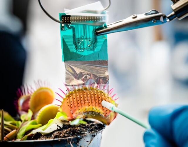
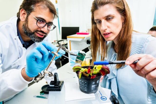
    Human beings and other animals send electrical signals via the central nervous system. The Venus flytrap, which lacks such a nervous system, also sends rapid electrical impulses, which are generated in response to touch or stress. It's how the plant traps its prey to feed. Now scientists have developed a bioelectronic device to better understand the Venus flytrap's complex signaling mechanism by mapping how those signals propagate, according to a recent paper published in the journal Science Advances.


    “We can now say with certainty that the electrical signal originates in the sensory hairs of the Venus flytrap," said co-author Eleni Stavrinidou of Linköping University in Sweden. "With our technology, we can also see that the signal mainly spreads radially from the hair, without any clear direction."


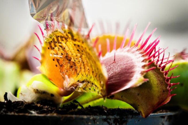
    As we've reported previously, the Venus flytrap attracts its prey with a pleasing fruity scent. When an insect lands on a leaf, it stimulates the highly sensitive trigger hairs that line the leaf. When the pressure becomes strong enough to bend those hairs, the plant will snap its leaves shut and trap the insect inside. Long cilia grab and hold the insect in place, much like fingers, as the plant begins to secrete digestive juices. The insect is digested slowly over five to 12 days, after which the trap reopens, releasing the dried-out husk of the insect into the wind.


    In 2016, Rainer Hedrich, a biophysicist at Julius-Maximilians-Universität Würzburg in Bavaria, Germany, led the team who discovered that the Venus flytrap could actually "count" the number of times something touches its hair-lined leaves—an ability that helps the plant distinguish between the presence of prey and a small nut or stone, or even a dead insect. The plant detects the first "action potential" but doesn't snap shut right away, waiting until a second zap confirms the presence of actual prey, at which point the trap closes. But the Venus flytrap doesn't close all the way and produce digestive enzymes to consume the prey until the hairs are triggered three more times (for a total of five stimuli).


    flytrap3-640x500.jpg

    The measuring device consists of a film with many electrodes in it, so thin that it can follow the curvature of the plant's lobes.
    Thor Balkhed/Linköping University


    In 2020, Japanese scientists genetically altered a Venus flytrap to gain important clues about how the plant's short-term "memory" works. They introduced a gene for a calcium sensor protein called GCaMP6, which glows green whenever it binds to calcium. That green fluorescence allowed the team to visually track the changes in calcium concentrations in response to stimulating the plant's sensitive hairs with a needle. They concluded that the waxing and waning of calcium concentrations in the leaf cells really do seem to serve as a kind of short-term memory for the Venus flytrap, though precisely how calcium concentrations work with the plant's electrical network remains unclear.


    However, a mutant Venus flytrap dubbed Dyscalculia (DYSC) does not close in response to two sensory stimuli, nor does it process its prey in response to additional stimuli. It has somehow "forgotten' how to count. Earlier this year, Hedrich and his team found that the mutation did not seem to affect either the action potential or the underlying calcium signal in the first two-count stage of the process. The action potentials fire, yet the trap doesn't snap shut, suggesting that the touch-activation of calcium signaling is being suppressed.


    Despite these recent advances, Stavrinidou et al. note that a scientific understanding of the underlying mechanisms and the precise relationship of action potentials with plant function is still incomplete, in part because it is challenging to accomplish high-resolution mapping of the signal. So they turned to bioelectronics, specifically noninvasive electrophysiological recording devices often used to study mammalian systems. The team adapted this approach to the Venus flytrap, building a multi-electrode micro-array made up of a very thin film embedded with electrodes.


    The film wrapped around the outside of the lobes of the Venus flytrap, and the researchers would then poke one of the plant's sensory hairs and measure the resulting signal in the lobe. They also filmed how the plant moved so they could see if the closure of the Venus flytrap correlated with the electrical signal. Prior measurement attempts had relied on a single measuring point; Stavrinidou et al. used around 30 electrodes for their signal measurements, which helped them pinpoint the origin of the signal and the direction in which it spread. They were also able to investigate the role of ions in the spread of the electrical signal, thanks to pharmacological treatments involving ion blockers.


    flytrap2-640x426.jpg

    Abdul Manan Dar and Eleni Stavrinidou at Linköping University, Sweden, demonstrate how the
    multi-electrode array technology can be used to examine the emergence and propagation of the
    electrical signal in a Venus flytrap.
    Thor Balkhed/Linköping University


    They found that the electrical signal starts in the plant's sensory hairs and then spreads radially outward with no clear preferred direction. And sometimes the signals were spontaneous, originating in sensory hairs that had not been stimulated. They don't yet know why this happens or what function this might serve. As for the ion experiment, the team didn't observe any significant impact in terms of how fast the signals spread when ion blockers were in play.


    “One of the most important aspects of this study is that we show that bioelectronic technologies, which are extensively used in biomedical research, can be applied to plant physiology research as well, therefore opening possibilities for new discoveries," said Stavrinidou. "There is currently a great need for developing plants that are more stress resistant for us to be able to grow food and have healthy forests also in the future. That’s why it’s important that we understand how plants respond to stress, and I think that this new technology may contribute in this area of research.”


    DOI: Science Advances, 2023. 10.1126/sciadv.adh4443  (About DOIs).


    Source


    User Feedback

    Recommended Comments

    There are no comments to display.


    Join the conversation

    You can post now and register later. If you have an account, sign in now to post with your account.
    Note: Your post will require moderator approval before it will be visible.

    Guest
    Add a comment...

    ×   Pasted as rich text.   Paste as plain text instead

      Only 75 emoji are allowed.

    ×   Your link has been automatically embedded.   Display as a link instead

    ×   Your previous content has been restored.   Clear editor

    ×   You cannot paste images directly. Upload or insert images from URL.


  • Recently Browsing   0 members

    • No registered users viewing this page.
×
×
  • Create New...