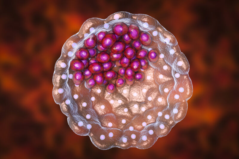
Scientists set a new record for growing primate embryos outside the womb, as reported in the May issue of the journal Cell. For the first time, monkey embryos were cultivated in a lab for 25 days post-fertilization, achieving key developmental landmarks never before observed in culture, including the start of organ development. The ability to track these processes in the lab might be an important step toward understanding congenital birth defects and organ development in humans.
Understanding development
The early stages of animal development, often referred to as embryogenesis, encompass the transition from a seemingly unremarkable clump of cells to a complex and compartmentalized organism. At the conclusion of embryogenesis, cells have started the march toward specialization, and organ systems have begun to form. In mammals, this is a process that usually happens in the comfort and privacy of the uterus, making it difficult to observe, even with the advent of advanced imaging. And it’s difficult to experiment with factors that might influence development.
All of this has led developmental biologists to search for ways to get this process to occur in a culture dish, bypassing these limitations. Studying human embryogenesis is restricted due to ethical and legal considerations. While the specific guidelines may vary from country to country, the outcome is a nearly global prohibition on lab-maintained human embryos past 14 days—before the progenitor of the nervous system forms. This detail is of particular medical relevance, as irregularities during nervous system formation can result in a range of conditions affecting the spine, spinal cord, and brain, including spina bifida.
Most of our understanding of this process comes from studies of mouse and chicken embryos.
But even the mouse is more than a hop, skip, and jump away from humans on the tree of life; while we're remarkably similar in some respects, there are notable differences in developmental timing and gene activity, and even the physical arrangement of the embryo is different.
Five more days
Two independent groups of researchers, publishing back-to-back papers, have tried to bridge this gap by developing a primate system for studying embryogenesis. Using fertilized embryos from a macaque species (Macaca fascicularis), both groups were able to cultivate primate embryos to 25 days post-fertilization. This surpasses the previous record of 20 days, allowing researchers to witness more advanced stages of organ development.
Each group developed a distinct technique, but both were able to get embryos that more closely resembled their in utero counterparts than ever before. One of the keys to achieving this was promoting three-dimensional growth of the embryo. Past efforts had relied on two-dimensional culture conditions, which were plagued by tissue overgrowth and ultimately embryo collapse by day 20. To address this, the team led by Hongmei Wang optimized culture conditions in a liquid suspension, whereas Tao Tan’s group used mechanical supports. Both approaches seemed to facilitate proper three-dimensional growth.
Survival of the embryos to day 25 ranged from approximately 20 to 40 percent, depending on the study and particular metric used. That might not exactly be an efficient, high-throughput success, but it stands as a significant improvement over earlier techniques. Importantly, the embryos surviving to 25 days had many of the hallmarks of an in utero embryo, such as the three embryonic tissue layers ultimately responsible for the formation of the fetus (the endoderm or gut lining, the ectoderm that forms skin and nerves, and mesoderm, which is everything else). The researchers also documented evidence of further organogenesis and formation of the neural tube, which goes on to form the brain and spinal cord.
This all makes for a very promising step toward a tractable system to study primate embryogenesis, but the techniques are far from perfect. Currently, the embryos don’t survive past 25 days in either set of conditions. However, both groups have suggested that an optimized mix of growth and signaling molecules, combined with more advanced mechanical supports, might better mimic conditions in the womb and sustain embryos into even later stages of development.
Cell, 2023. DOIs: 10.1016/j.cell.2023.04.019, 10.1016/j.cell.2023.04.020
Lita Kacsoh has a masters in public health from Emory University and a PhD in molecular biology from Dartmouth Medical School. When she isn't writing, she can probably be found reading or drinking coffee.


3175x175(CURRENT).thumb.jpg.b05acc060982b36f5891ba728e6d953c.jpg)
Recommended Comments
There are no comments to display.
Join the conversation
You can post now and register later. If you have an account, sign in now to post with your account.
Note: Your post will require moderator approval before it will be visible.