
Imaging techniques such as computed tomography, magnetic resonance imaging, positron emission tomography and ultrasound have become indispensable in the medical world. Each method not only opens unique insights into people's insides, but also allows physicians to draw conclusions about defects or functional processes in the human body.
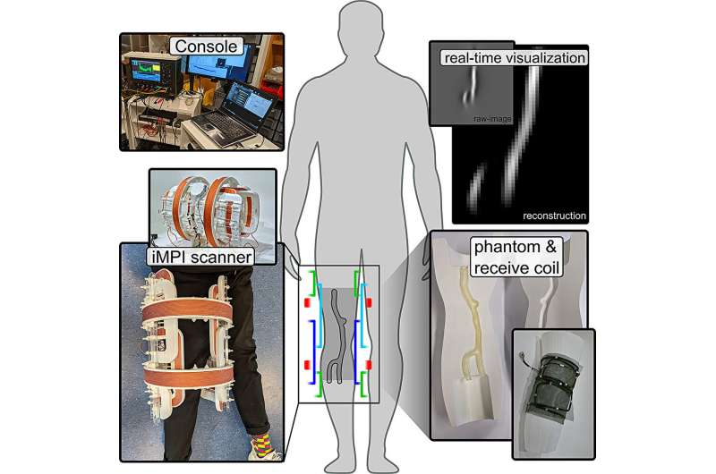
A team of physicists and medical doctors from the Julius-Maximilians-Universität Würzburg (JMU) has now succeeded in making another—and radiation-free—imaging technology ready for use on humans. It's called magnetic particle imaging (MPI). With the portable scanner they developed, it is possible, among other things, to visualize dynamic processes in the human body, such as blood flow.
Professor Volker Behr and Dr. Patrick Vogel from the University's Institute of Physics are responsible for this study, and they have now published their results in the journal Scientific Reports.
A sensitive and fast alternative
Magnetic particle imaging is a technique based, as the name suggests, on the direct visualization of magnetic nanoparticles. Such nanoparticles do not occur naturally in the human body and must be administered as markers. "As with positron emission tomography, which relies on the administration of radioactive substances as markers, this method has the great advantage of being sensitive and fast without 'seeing' interfering background signals from tissue or bone," explains Volker Behr.
MPI is not based on the detection of gamma rays from a radioactive marker like positron emission tomography, but on the response signal of the magnetic nanoparticles to magnetic fields that change over time. "In this process, the magnetization of nanoparticles is specifically manipulated with the help of external magnetic fields, whereby not only their presence but also their spatial position in the human body can be detected," says physicist Patrick Vogel, first author of the publication.
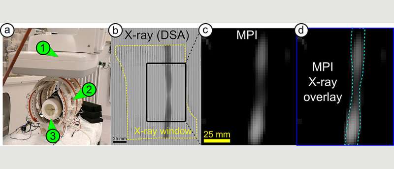
The iMPI scanner (left) provides new insights into the human body. Here you can see a constriction in a blood vessel—recorded with conventional X-rays (b), with the scanner (c) and in a combination of both techniques (d). Credit: Patrick Vogel / Stefan Herz
A small scanner for big insights
The MPI idea is not new. As early as 2005, the Philips company was able to show the first images of this novel approach in a small demonstrator, which, however, could only take samples a few centimeters in size. And the development of devices suitable for examining humans proved more difficult than expected, leading to large, heavy and expensive constructions.
In 2018, the team led by Professor Volker Behr and Patrick Vogel found a new way to implement the complex magnetic fields required for imaging in a much smaller design. In a multi-year research project, the scientists succeeded in implementing the novel concept in an MPI scanner (interventional Magnetic Particle Imaging—iMPI) specifically designed for intervention.
"Our iMPI scanner is so small and light that you can take it almost anywhere," Vogel explains. The authors impressively demonstrate this mobility of the scanner in a simultaneous real-time measurement in comparison with a special X-ray device, which is the standard device in angiography in university hospitals.
The team led by Professor Thorsten Bley and Dr. Stefan Herz of the Interventional Radiology Department of the Würzburg University Hospital, which accompanied this project from the beginning, carried out the measurements on a realistic vascular phantom and evaluated the first images.
"This is a first important step towards radiation-free intervention. MPI has the potential to change this field for good," said Dr. Stefan Herz, senior author of the publication.
In addition to further measurements with the iMPI device, the two physicists are now working to further develop their scanner. The main goal is to improve the image quality.
- digitaldon2 and Karlston
-
 2
2
Recommended Comments
There are no comments to display.
Join the conversation
You can post now and register later. If you have an account, sign in now to post with your account.
Note: Your post will require moderator approval before it will be visible.