
A team of neuroscientists and physiologists at the University of Cambridge, working with a pair of colleagues from the Wellcome Trust-MRC Institute of Metabolic Science at Addenbrooke's Hospital, has found that MRI scans can reveal the parts of the brain that are involved in responding to fatty foods. In their paper published in The Journal of Neuroscience, the group describes how they tested milkshakes with different amounts of fat, then asked volunteers to taste them while undergoing brain scans, and what they found by doing so.
Prior research has shown that one of the factors involved in the obesity epidemic in many Western countries is a preference for fatty foods. In this new effort, the team in the U.K. sought to learn which parts of the brain are involved in such a preference—a finding that might lead to therapies directed at those brain parts to help reduce peoples' response to fatty foods.
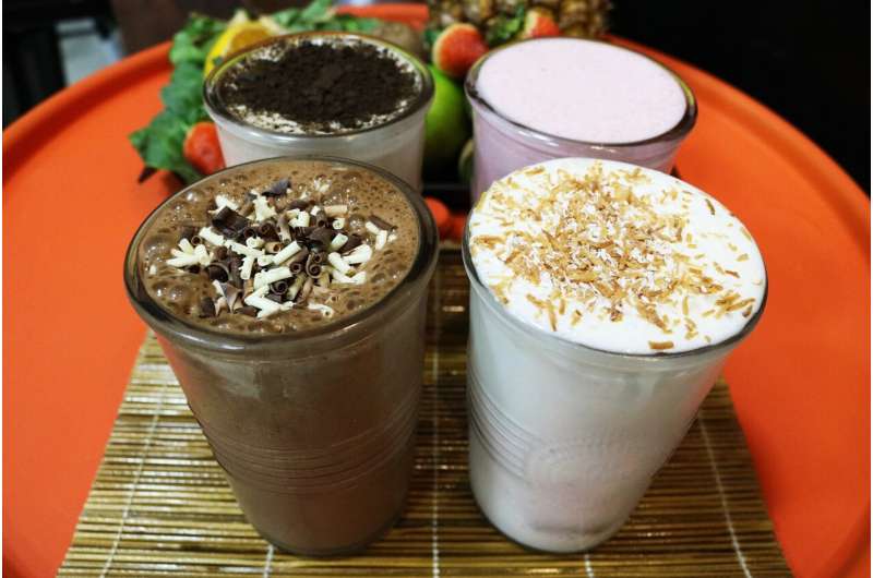
One of the reasons people like ice cream is its texture—and its texture is partly due to the amount of fat it contains. Prior research has shown that people respond to both the taste of the ice cream and the rich physical sensations that are felt as the ice cream is eaten, particularly when licked from atop a cone. To find out which parts of the brain are responsible for such reactions, the research team carried out a series of measurements.
The first measurement involved creating several milkshakes with varying levels of fat in them. Next, they used tongues extracted from pigs to measure the degree of friction that occurred when each of the milkshakes was licked.
They then asked 22 human volunteers to sample the same types of milkshakes, while also undergoing an MRI scan of the brain. After each taste, each volunteer was asked to estimate how much they would pay for a full glass of the milkshake as a means of measuring how much each of the volunteers liked the samples they were given.
In studying the brain scans, the researchers found that the orbitofrontal cortex (OFC) lit up when the volunteers were taste-testing. They also found that the amount it lit up corresponded with the amount a volunteer was willing to pay for a shake after a taste-test. The study concluded by inviting all the volunteers to a free meal—curry dishes were given with different amounts of fat in them. The researchers found that those volunteers whose OFCs lit up the most, tended to eat more of the curry that had the most fat in it.
Recommended Comments
There are no comments to display.
Join the conversation
You can post now and register later. If you have an account, sign in now to post with your account.
Note: Your post will require moderator approval before it will be visible.